

The patellar tendon connects the kneecap to the shin at the front of the knee. Patellar tendinopathy is most common in young people (teenagers and young adults) and in jumping sports — basketball, volleyball, jumping events — and less so in football, weightlifting or cycling.
It is usually caused by overload in training and competition; less often by a direct blow. Risk factors include height, higher body weight, skeletal immaturity and reduced flexibility of the quadriceps and hamstrings; among external factors, hard training surfaces.
Biomechanically, the greatest load comes not on take-off but on landing, when the tendon undergoes an eccentric contraction — contracting while lengthening.
The four clinical stages
- Pain after training.
- Pain during and after training, without affecting performance.
- Pain during and after training that affects performance.
- Rupture of the tendon.
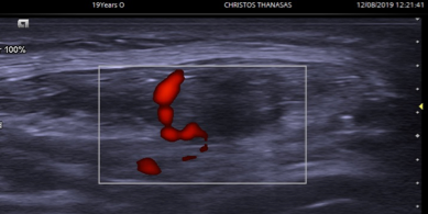
The pain typically sits where the tendon leaves the kneecap, centrally or to the outside, and peaks during jumps and sprints. In advanced stages it is present in daily life too. Besides a clinical examination, an ultrasound scan and possibly an MRI are needed.
Treatment
If not treated in the first weeks, symptoms usually become chronic. In chronic cases the tendon shows thickening, micro-tears, abnormal blood vessels and bone spurs at the pole of the kneecap. Patellar tendinopathy is not easy to treat and needs a team: orthopaedic surgeon, rehabilitation physician, physiotherapist and trainer.
- Reducing the intensity, volume and type of training.
- Physiotherapy and isometric contractions to reduce symptoms.
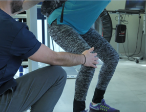
- An exercise programme for quadriceps and hamstring flexibility, with eccentric work such as Heavy Slow Resistance training, isoinertial exercise and the Alfredson protocol.
- Ultrasound-guided injections: PRP, high-volume, electrolysis.
These conservative treatments are the most effective, and the right combination often provides a lasting solution.
When conservative treatment fails
Until recently the usual answer was open surgery. Today we can achieve a better result, in less time, with Sonosurgery: removing the abnormal vascular and nerve tissue, bone spurs, calcifications and bursae that cause the symptoms.
The procedure
It is done in hospital under local or general anaesthetic. The diseased area is located with ultrasound and two tiny incisions are made. Using a camera and micro-instruments the diseased tissue is cleared (arthroscopy) without harming the healthy tendon; the result is confirmed on ultrasound. It takes less than an hour.
Advantages
- A definitive treatment.
- High precision; the healthy part of the tendon is untouched.
- The tendon is not detached or cut — less discomfort and fewer complications.
- No hospital stay.
- Faster recovery than open surgery.
Before the procedure
- You may eat up to three hours before.
- Continue your medication, except anti-inflammatories (stop three days before) and anticoagulants (only if your doctor agrees).
- Arrange not to drive home.
After the procedure
- Painkillers if needed; antibiotics only if you are at high risk of infection.
- A bandage for 48 hours; walking with crutches a few hours later, for 5–7 days.
- You can shower after 48 hours, using a waterproof adhesive dressing.
- Two stitches removed at 2 weeks; a physiotherapy and exercise programme.
- Driving in 1–2 weeks; office work in 2–3 days; physical work in six weeks.
- Gradual return to sport after six weeks.
This information is for general guidance and does not replace a medical consultation. The right treatment is always decided individually, after clinical assessment.


