In brief
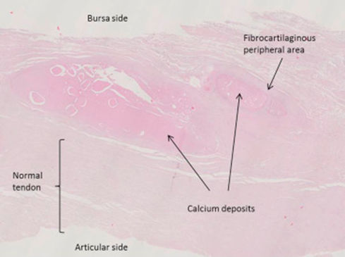
- Calcium collects inside the shoulder tendons, most often in supraspinatus near where it attaches to the humerus.
- The deposit is made of hydroxyapatite crystals — the same mineral normally found in bone.
- Mechanism: "cell metaplasia". Fibroblasts turn into chondroblasts and osteoblasts and lay down calcium, probably in response to increased load.
- It affects people aged 30–60, slightly more often women. Diabetes, thyroid disease and rheumatological conditions raise the risk; a genetic predisposition is possible.
- It causes pain on movement and often at rest, which can be intense, along with crepitus, catching and restricted shoulder movement.
The natural course of the disease
Pre-calcific stage
Normal fibroblasts turn into chondroblasts and osteoblasts. There are no symptoms yet.
Calcific stage
- Formative phase: calcium accumulates; it may cause symptoms.
- Resting phase: the deposit stabilises; in many cases it is troublesome.
- Resorptive phase: the calcium is absorbed by phagocytosis; this is the most painful phase.
Post-calcific stage
The body tries to restore the normal structure of the tendon.
In many patients the resting phase lasts for years and never moves on to resorption.
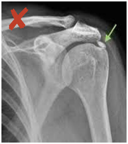
X-ray classification (Gärtner & Heyer)
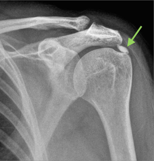
- Type I: well-defined, dense — formative phase.
- Type II: smooth or sharp outline, dense, with radiolucent areas — resting phase.
- Type III: translucent, cloudy, poorly defined — resorptive phase.
Ultrasound classification (Farin)
In clinical practice, ultrasound gives the most reliable information about the deposit’s consistency:
- Type A: hyperechoic with a strong acoustic shadow.
- Type B: hyperechoic with a faint acoustic shadow.
- Type C: hyperechoic with no acoustic shadow.
What this means for treatment
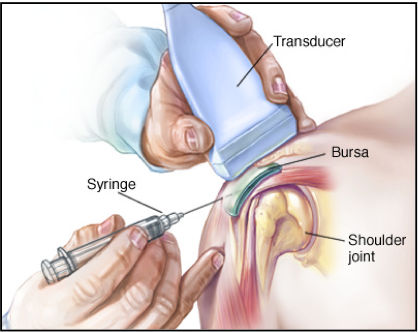
The classification lets us predict how well barbotage will work. When the deposit is soft or moderately hard, an excellent or very good result is expected; when it is hard, the result is likely to be moderate or poor. The best indication for barbotage is a Farin B or C, Gärtner/Heyer II or III deposit.
Consistency is an important predictor, but not the only one. Your doctor will also weigh the deposit’s position and size and any associated problem (e.g. a partial-thickness tear, the subacromial space) before recommending treatment.
This information is for general guidance and does not replace a medical consultation. The right treatment is always decided individually, after clinical assessment.